File:Fig6 SECM.jpg
Jump to navigation
Jump to search

Size of this preview: 800 × 366 pixels. Other resolutions: 320 × 146 pixels | 640 × 293 pixels | 1,183 × 541 pixels.
Original file (1,183 × 541 pixels, file size: 48 KB, MIME type: image/jpeg)
Summary
| Description |
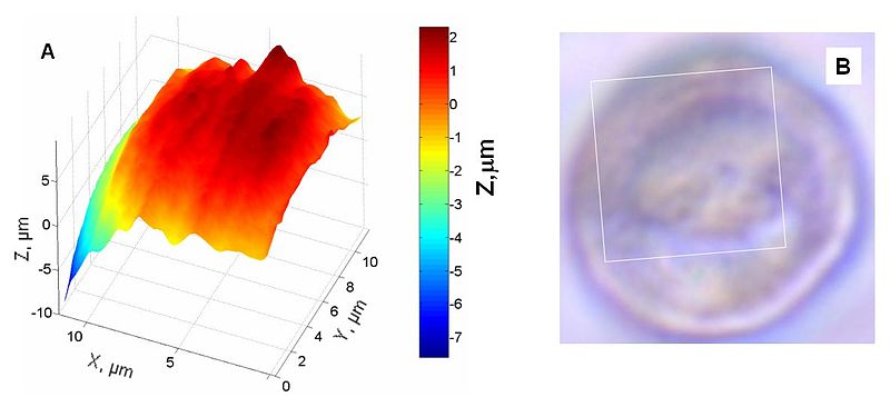
English: Fig. 6. Substrate imaging (constant current mode). (A) SECM image of a portion (10 µm × 10 µm) of a human breast cell using a 120 nm radius tip. (B) Optical micrograph of the same cell showing the SECM image area delimited by a white square. |
| Date | |
| Source | Own work |
| Author | Francois laforge |
Licensing
I, the copyright holder of this work, hereby publish it under the following license:
This file is licensed under the Creative Commons Attribution-Share Alike 3.0 Unported license.
- You are free:
- to share – to copy, distribute and transmit the work
- to remix – to adapt the work
- Under the following conditions:
- attribution – You must give appropriate credit, provide a link to the license, and indicate if changes were made. You may do so in any reasonable manner, but not in any way that suggests the licensor endorses you or your use.
- share alike – If you remix, transform, or build upon the material, you must distribute your contributions under the same or compatible license as the original.

File history
Click on a date/time to view the file as it appeared at that time.
| Date/Time | Thumbnail | Dimensions | User | Comment | |
|---|---|---|---|---|---|
| current | 23:16, 13 November 2011 | | 1,183 × 541 (48 KB) | wikimediacommons>Francoislaforge |
File usage
There are no pages that use this file.